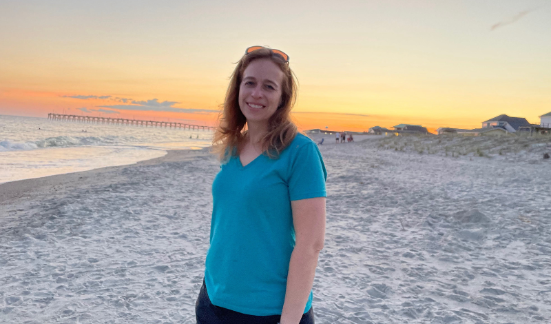
Advances in medical technology have led to improved detection and outcomes for lung cancer patients like Julie Kelly. Robotic bronchoscopy is one such innovation that allowed doctors to identify and diagnose her disease in its early stages. This minimally invasive procedure uses a robotic arm guided by a surgeon to biopsy hard-to-reach lung tumors. When a routine mammogram led to the discovery of an unusual lung mass, robotic bronchoscopy provided critical insights that revealed Julie’s cancer.
In January 2022, then 42-year-old Julie Kelly went in for a routine mammogram. She had no symptoms of any kind and was feeling well. The films from her mammogram were clear, but the radiologist did notice a calcified blood vessel. At the time, physicians had started to flag things like this as incidental findings because they are closely correlated to atherosclerosis elsewhere in the body, including coronary arteries. This information led the doctors to encourage Julie to follow up with her primary care physician (PCP). At the time, no one, Julie included, was particularly concerned, and off she went.
When she saw her PCP in March, they spent a lot of time discussing her family medical history and any symptoms she might be experiencing; there were none. The doctor wanted Julie to undergo a heart CT scan, an exploration of her calcium score, and testing on the condition of her coronary arteries. When testing came back indicating a 3 cm mass in her lung, Julie was certain there had to be a mistake. Together with her doctor, they agreed to do nothing for one month and then re-scan and reassess.
The Growth That Wasn’t Supposed to Be There
One month later, imaging confirmed that the mass was still there. Despite the “good” news that it had not grown (nor shrunk, for that matter), Julie was referred to a pulmonologist to begin the process of assessing the situation and determining a plan. Upon looking at the films, the pulmonologist stated simply,
“That is not supposed to be there.”
Julie inquired whether the mass was a holdover from her recent bout of COVID-19. It was not. They further discussed risk factors. The only thing they came up with was exposure to secondhand smoke from her grandfather. Since he had passed away when Julie was just eight years old, it seemed highly unlikely that this exposure played a role. By this time, despite no one having made even a suggestion that this might be lung cancer, Julie was growing increasingly suspicious of the mysterious mass in her chest. So, too, was the pulmonologist.
Bronchoscopy Results Inclusive
Directly after that appointment, Julie was scheduled for an urgent PET scan. It was a huge relief that nothing else lit up, bringing a modicum of comfort in an increasingly uncomfortable situation. However, very soon after this scan, Julie underwent a bronchoscopy. While they were able to see that there had been no further tumor growth, doctors were unable to reach the tumor. The results: inconclusive.
Julie recalled what came next:
“By all accounts, it would have been appropriate for the pulmonologist to take a wait-and-see attitude. Typically, a situation like mine would be monitored every three months. But he recommended that we take a look again the following month. A CT scan in June confirmed that the lump – although it had not grown – was still there. In hopes of arriving at a diagnosis, I sought a second opinion which, to my great disappointment, proved worthless. The physician did not have my films and was reliant on the reports alone. I recall him commenting that my lump was something people die ‘with’, not ‘from’. As you can imagine, that was not as encouraging as one might think.”
Robotic Bronchoscopy Detects Lung Cancer
Still searching for answers – and aware that no one has indicated that this could be cancer – Julie went to see a thoracic surgeon. She was relieved and pleased when it became clear that he had done his due diligence and familiarized himself with every element of her journey thus far. He agreed that the lump should be removed and referred her to a physician who specializes in robotic bronchoscopy. This procedure, like a traditional bronchoscopy, is minimally invasive and allows doctors to biopsy nodules in the lungs.
The difference is that in robotic bronchoscopy, the doctor uses a controller at a console to operate a robotic arm. The most significant benefit is that robotic-assistant bronchoscopy can access lung nodules that previously required more invasive biopsy techniques or even surgery. Because it allows pulmonologists to reach tiny distal airways in the lungs, they can detect lung cancer in an earlier stage when it is easier to treat and potentially cure. Following this procedure, Julie got her diagnosis: non-small cell lung cancer, stage 1B.
Following diagnosis, Julie was scheduled for a lower left lobectomy to remove the tumor and surrounding tissue. The surgery – like the bronchoscopy – was performed robotically requiring a chest tube in the front along with 4 or 5 incisions through Julie’s back. Thankfully, the physician also sent out a specimen for biomarker testing. This test determined that Julie carried the EGFR mutation. Due, in part, to some discrepancies in lab reporting, her medical team was not in total agreement on the best protocol to pursue. Julie decided at that point to actively advocate for herself:
“The surgeon was confident that it was early stage and that he’d cured me, saying I only needed a scan every 6 months. I had to ask for an oncologist referral. When I met with the oncologist, she misread my biomarker report as well as my lab work, which had some inconsistencies in the report. My sister and I read up on the biomarker report and learned that I should be eligible for targeted therapy. So, I got a second opinion again at Duke.
This time, the second opinion was the key to peace of mind for a surveillance plan and treatment. The oncologist there has been involved in EGFR research. He requested my samples to confirm my stage as IB due to visceral pleural invasion. He then confirmed that I was eligible for targeted therapy, and I started treatment about 7 weeks after surgery. I now have chest and abdomen CT scans every 3 months and a brain MRI every 6 months.”
Robotic Bronchoscopy and Robotic Surgery Key to NED
Now, a little over a year from when this all began, Julie remains on medication and is thrilled to be NED (no evidence of disease). She will continue to be monitored every 3 months with CTs of her chest and stomach as well as brain MRIs every six months.
“Support groups made a huge difference for me in the whole process because people sharing their stories and the difference in care help you know what to ask for and what the standard of care can be across different centers and hospitals. It was a support group member who informed me of robotic bronchoscopy, and I also learned from support groups about the more frequent surveillance.”
When this all began, Julie didn’t quite believe the medical team when they, at the beginning of this journey, assured her she would be able to travel with her husband and three children on a long-planned and highly anticipated trip to Disney World. Thanks to early detection and amazing advances in lung cancer research, the Kelly family not only went to Disney but had the time of their lives…and had a lot to be grateful for.
Now in remission, Julie’s story illustrates how robotic bronchoscopy is transforming lung cancer care. This precise, state-of-the-art technique gave Julie’s medical team the information they needed to move quickly from diagnosis to successful treatment. As pioneering procedures like robotic bronchoscopy continue to advance early intervention for lung cancer, more patients can hope to share in Julie’s positive outcome. Ongoing targeted therapies and monitoring have kept her cancer-free, allowing Julie and her family to enjoy the life they deserve. The promise of life-changing innovations like robotic bronchoscopy offers renewed optimism in the fight against this disease.

